Introduction
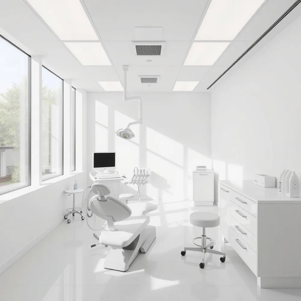
Cone‑Beam Computed Tomography (CBCT) captures a fast, low‑dose, three‑dimensional scan of the teeth, jaws, nerves and surrounding structures. This modern imaging replaces multiple 2‑D X‑rays with a single, detailed view, allowing dentists to diagnose hidden pathology, assess bone quality and evaluate vital anatomy with unprecedented clarity. In today’s dental practices CBCT drives precise implant placement, orthodontic planning and endodontic treatment, reducing complications and improving predictability. For patients, the technology means fewer appointments, lower radiation exposure, clearer explanations of their oral health and a more personalized, comfortable care experience.
Enhanced Diagnostic Accuracy with CBCT
![]() Cone‑Beam Computed Tomography (CBCT) delivers true three‑dimensional visualization of teeth, bone, nerves, and soft tissues, giving dentists a complete view that traditional two‑dimensional X‑rays cannot provide. This high‑resolution 3‑D picture reveals hidden pathologies—such as cysts, tumors, early bone loss, and temporomandibular joint (TMJ) disorders—that might otherwise go unnoticed. It also allows precise assessment of root morphology, cracked teeth, and complex canal anatomy, helping endodontists locate hidden canals and determine the full extent of a fracture.
Cone‑Beam Computed Tomography (CBCT) delivers true three‑dimensional visualization of teeth, bone, nerves, and soft tissues, giving dentists a complete view that traditional two‑dimensional X‑rays cannot provide. This high‑resolution 3‑D picture reveals hidden pathologies—such as cysts, tumors, early bone loss, and temporomandibular joint (TMJ) disorders—that might otherwise go unnoticed. It also allows precise assessment of root morphology, cracked teeth, and complex canal anatomy, helping endodontists locate hidden canals and determine the full extent of a fracture.
What are the benefits of a CBCT scan? A CBCT scan produces a detailed 3‑D image of the maxillofacial region, enabling faster, more accurate diagnoses and treatment planning. Because the entire area is captured in a single low‑dose exposure, patients receive less radiation than they would from multiple conventional X‑rays, and the scan is completed in seconds. The data can be used to design custom surgical guides, virtual models, and orthodontic appliances, which reduces guesswork and improves clinical outcomes.
What are the advantages of using three‑dimensional digital imaging in dental diagnostics? Three‑dimensional imaging offers a clearer, more complete picture of oral health. By reconstructing dozens of images into a 3‑D model, dentists can evaluate tooth position, bone density, and nerve pathways with millimeter precision, leading to safer implant placement, more predictable orthodontic movement, and better overall treatment planning.
Will CBCT show a cracked tooth? Yes. While a superficial crack may be visible during a clinical exam, CBCT can determine the depth and orientation of the fracture and assess any associated pulp‑periapical or periodontal involvement, guiding appropriate restorative or surgical intervention.
Overall, CBCT enhances diagnostic confidence, reduces radiation exposure, and supports personalized, patient‑centered care.
Precision Implant Planning and Surgical Guidance
![]() Cone‑Beam Computed Tomography (CBCT) delivers high‑resolution three‑dimensional images that enable clinicians to measure bone volume, density, and quality with millimeter precision. This accurate assessment is essential for selecting the appropriate implant size and angulation, and it significantly reduces the risk of nerve injury or sinus perforation by clearly visualizing vital structures such as the inferior alveolar nerve and the maxillary sinus. The detailed 3‑D data also serve as the foundation for creating virtual surgical guides and computer‑aided implant placement. By integrating CBCT scans with CAD/CAM software, dentists can design patient‑specific guides that direct drilling and implant insertion, streamlining the procedure, shortening chair‑time, and improving predictability of outcomes.
Cone‑Beam Computed Tomography (CBCT) delivers high‑resolution three‑dimensional images that enable clinicians to measure bone volume, density, and quality with millimeter precision. This accurate assessment is essential for selecting the appropriate implant size and angulation, and it significantly reduces the risk of nerve injury or sinus perforation by clearly visualizing vital structures such as the inferior alveolar nerve and the maxillary sinus. The detailed 3‑D data also serve as the foundation for creating virtual surgical guides and computer‑aided implant placement. By integrating CBCT scans with CAD/CAM software, dentists can design patient‑specific guides that direct drilling and implant insertion, streamlining the procedure, shortening chair‑time, and improving predictability of outcomes.
What are the advantages of 3D imaging? 3‑D imaging provides a concise, volumetric view of oral anatomy, making diagnosis faster and easier to interpret. It enhances communication among clinicians and with patients, boosting confidence and informed consent. The technology supports precise treatment planning, reduces the need for multiple radiographs, and lowers overall radiation exposure when ALARA protocols are followed. Ultimately, 3‑D imaging improves clinical efficiency, minimizes complications, and delivers higher‑quality, patient‑centered dental care.
Orthodontic and Orthognathic Treatment Optimization
![]() Cone‑Beam Computed Tomography (CBCT) has become the cornerstone of modern orthodontic and orthognathic planning. Its three‑dimensional visualization lets clinicians evaluate tooth positioning, root orientation, and jaw relationships with millimeter precision, eliminating the superimposition that limits traditional 2‑D X‑rays. By capturing the entire maxillofacial skeleton, CBCT reveals skeletal asymmetry and airway dimensions, enabling accurate diagnosis of complex malocclusions and informing decisions about surgical versus non‑surgical correction. The detailed anatomy also guides the design of customized orthodontic appliances and clear‑aligner therapy—digital models derived from CBCT data ensure that forces are applied exactly where needed, shortening treatment time and improving predictability.
Cone‑Beam Computed Tomography (CBCT) has become the cornerstone of modern orthodontic and orthognathic planning. Its three‑dimensional visualization lets clinicians evaluate tooth positioning, root orientation, and jaw relationships with millimeter precision, eliminating the superimposition that limits traditional 2‑D X‑rays. By capturing the entire maxillofacial skeleton, CBCT reveals skeletal asymmetry and airway dimensions, enabling accurate diagnosis of complex malocclusions and informing decisions about surgical versus non‑surgical correction. The detailed anatomy also guides the design of customized orthodontic appliances and clear‑aligner therapy—digital models derived from CBCT data ensure that forces are applied exactly where needed, shortening treatment time and improving predictability.
How has medical imaging improved healthcare? Medical imaging dramatically increases diagnostic accuracy by exposing hidden anatomical and functional details, allowing earlier disease detection and more confident treatment planning. In dentistry, CBCT provides high‑resolution 3‑D data that refine risk assessment, reduce invasive guesswork, and support personalized, data‑driven care, ultimately enhancing patient outcomes while minimizing radiation exposure and procedural costs.
Endodontic Excellence and Complex Anatomy
![]() Cone‑Beam Computed Tomography (CBCT) delivers high‑resolution, three‑dimensional images that reveal the intricate morphology of root canals, including hidden accessory canals that are invisible on traditional 2‑D radiographs. This detailed view enables endodontists to locate and negotiate complex canal systems with confidence, reducing the risk of missed anatomy that can lead to treatment failure. CBCT also excels at detecting vertical root fractures and subtle periapical pathology, such as early‑stage lesions or bone defects, by visualizing the tooth and surrounding bone in multiple planes. When surgery or retreatment is required, the three‑dimensional data can be integrated into guided endodontic procedures, allowing precise planning of access pathways, instrument trajectories, and surgical guides. This level of precision shortens chair‑time, improves healing outcomes, and enhances patient confidence by providing a clear visual explanation of the condition and proposed treatment.
Cone‑Beam Computed Tomography (CBCT) delivers high‑resolution, three‑dimensional images that reveal the intricate morphology of root canals, including hidden accessory canals that are invisible on traditional 2‑D radiographs. This detailed view enables endodontists to locate and negotiate complex canal systems with confidence, reducing the risk of missed anatomy that can lead to treatment failure. CBCT also excels at detecting vertical root fractures and subtle periapical pathology, such as early‑stage lesions or bone defects, by visualizing the tooth and surrounding bone in multiple planes. When surgery or retreatment is required, the three‑dimensional data can be integrated into guided endodontic procedures, allowing precise planning of access pathways, instrument trajectories, and surgical guides. This level of precision shortens chair‑time, improves healing outcomes, and enhances patient confidence by providing a clear visual explanation of the condition and proposed treatment.
Patient‑Centered Care, Education, and Insurance
![]() Cone‑Beam Computed Tomography (CBCT) creates three‑dimensional visualizations that dentists can display on a screen during the visit. By rotating the model and zooming in on nerves, bone density, and tooth roots, patients see exactly what the clinician sees. This visual aid demystifies the diagnosis, clarifies why a particular implant or orthodontic approach is needed, and builds trust, leading to informed consent and higher treatment acceptance.
Cone‑Beam Computed Tomography (CBCT) creates three‑dimensional visualizations that dentists can display on a screen during the visit. By rotating the model and zooming in on nerves, bone density, and tooth roots, patients see exactly what the clinician sees. This visual aid demystifies the diagnosis, clarifies why a particular implant or orthodontic approach is needed, and builds trust, leading to informed consent and higher treatment acceptance.
Because a single CBCT scan captures the entire maxillofacial region, it replaces multiple traditional radiographs. The technology’s focused, cone‑shaped beam delivers a radiation dose that is 10‑30 times lower than a conventional medical CT and comparable to a few panoramic X‑rays, adhering to the ALARA principle while still providing high‑resolution 3‑D data.
Insurance coverage for CBCT varies, but many Delta Dental PPO and Premier plans include it. The specific diagnostic codes D0365 (full‑arch mandibular) and D0366 (full‑arch maxillary, with or without cranium) are typically covered, limited to one scan per 12‑month period and subject to plan coinsurance and exclusions. Patients should verify benefits through the Delta Dental portal or by contacting the insurer, as coverage may differ by individual plan design.
Q: Does Delta Dental cover CT scans? A: Yes, Delta Dental’s PPO and Premier plans include coverage for cone‑beam CT (CBCT) scans. The specific diagnostic codes D0365 (full‑arch mandibular) and D0366 (full‑arch maxillary, with or without cranium) are covered. Scans are limited to one per 12‑month period and are subject to the plan’s overall limitations, exclusions, and any applicable coinsurance or out‑of‑pocket costs. Coverage can vary by individual plan design, so members should verify their benefits through the provider portal or by contacting Delta Dental. In short, most Delta Dental PPO/Premier policies do cover CBCT imaging, but you must check your specific plan for any additional requirements.
Conclusion
CBCT transforms treatment planning by delivering three‑dimensional views that (1) reveal hidden anatomy such as impacted teeth and root canals, (2) measure bone volume and density for implant placement, (3) map structures like the inferior alveolar nerve and sinus cavities, (4) evaluate skeletal relationships for orthodontic and orthognathic cases, and (5) monitor healing progress after surgery. The technology uses a focused cone‑beam, delivering lower radiation than conventional CT and meeting the ALARA principle, which safeguards patients while enhancing confidence. At Southern Boulevard Dental we invite you to ask about CBCT options so we can create a plan for your smile.