Introduction to Digital Dentistry and 3D Imaging
Understanding Digital Dentistry
Digital dentistry encompasses a range of advanced technologies including intraoral scanners, digital radiography, CAD/CAM systems, 3D printing, and artificial intelligence. These technologies collectively transform traditional dental practices by enabling more precise diagnostics, treatment planning, and restorative procedures.
Emergence and Benefits of Digital Imaging and 3D Scanning
Digital imaging technologies like cone beam computed tomography (CBCT) and 3D scanning provide detailed, three-dimensional views of teeth, gums, jawbones, and surrounding structures. This enhanced visualization supports early detection of dental issues, accurate assessment of bone density, nerve pathways, and oral anatomy, dramatically improving diagnostic accuracy and personalized treatment plans.
Impact on Clinical Workflows and Patient Experience
The integration of digital impressions and 3D imaging streamlines dental workflows by reducing the need for traditional molds, minimizing chair time, and allowing same-day restorations. Patients benefit from increased comfort, as digital scans eliminate the discomfort associated with conventional impression materials. Additionally, these technologies facilitate better communication, with realistic 3D models enhancing patient understanding and engagement in their dental care.
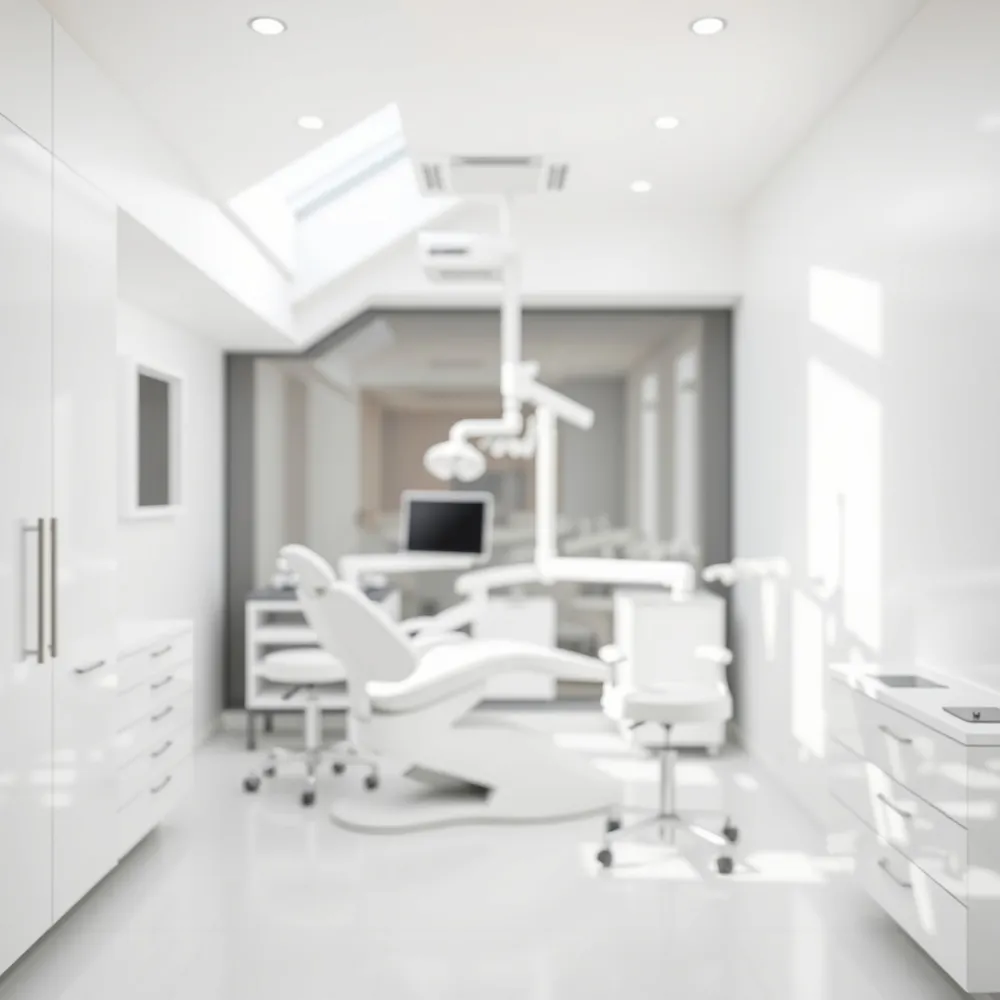
Understanding Digital Imaging and 3D Scanning in Modern Dentistry
![]()
What is digital imaging in dentistry?
Digital imaging in dentistry is a modern technique that uses digital sensors and specialized software to capture high-resolution, instant radiographic images. This approach replaces traditional film X-rays, reducing radiation exposure by up to 75% while providing detailed views of teeth, bones, and soft tissues that traditional methods cannot reveal. Digital imaging allows dentists to manipulate images in real time, securely store them, and share results quickly, enhancing both diagnosis and patient care. This technology streamlines clinical workflows and increases accuracy, improving overall dental treatment quality.
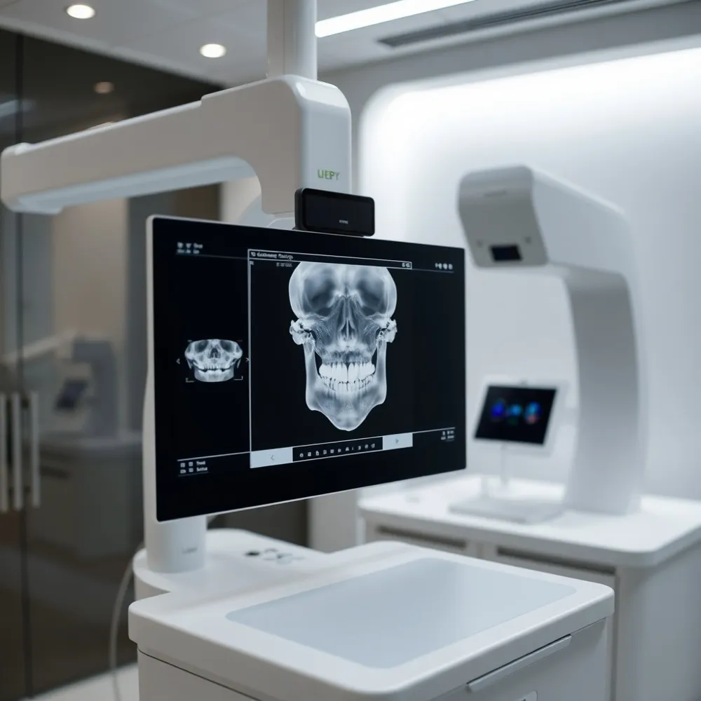
What is 3D imaging in dentistry?
Known as Cone Beam Computed Tomography (CBCT), 3D imaging produces three-dimensional views of oral structures. Unlike conventional two-dimensional X-rays, CBCT captures precise images of teeth, gums, jawbones, nerves, and sinuses. This detailed visualization supports critical procedures such as implant placement, root canal therapy, orthodontics, and oral surgery by allowing for meticulous treatment planning and reducing risks. Using 3D imaging ensures safer, more effective dental care with personalized treatment tailored to each patient's anatomy.
What is the difference between 3D digital scans and CBCT scans?
3D digital scans, or intraoral scans, use light-based technologies like lasers or structured light to create detailed surface images of the mouth. These scans are primarily used for digital impressions in restorative work, orthodontics (like Invisalign), and patient education. Conversely, CBCT scans employ low-dose X-rays to generate comprehensive, volumetric views of the internal bone and soft tissue structures. While intraoral scans focus on surface detail for appliances and restorations, CBCT provides indispensable insight into internal anatomy necessary for complex surgical planning.
How much radiation is involved in a 3D dental scan?
CBCT scans expose patients to a relatively low level of radiation, typically ranging from 0.003 to 1.073 millisieverts (mSv). A standard dental CBCT scan usually involves about 0.087 mSv, equating to roughly 11 days of natural background radiation—comparable or lower than other dental imaging methods. Dental professionals adhere to the ALARA principle ("As Low As Reasonably Achievable") to minimize patient exposure, especially in children. Consequently, 3D imaging balances exceptional diagnostic benefits with patient safety effectively.
Advantages over traditional X-rays
Digital imaging and 3D scans enhance diagnostic accuracy by providing detailed images that surpass standard flat X-rays. They allow immediate viewing and manipulation of images, reduce radiation doses significantly, and improve patient comfort by eliminating the need for uncomfortable molds or invasive procedures. Furthermore, these technologies support patient education with clear visual aids, enable faster treatment planning, and facilitate remote consultations via digital sharing, all contributing to improved patient outcomes and dental practice efficiency.
Advantages and Clinical Benefits of Digital Scanning and Imaging
![]()
What are the advantages of digital scanning in dentistry?
Digital scanning in dentistry provides a range of significant benefits that enhance both patient care and clinical outcomes. First and foremost, it offers exceptional accuracy and precision in dental restorations. Highly detailed, three-dimensional images of teeth, gums, and surrounding tissues allow dentists to create crowns, bridges, veneers, and other restorations with a superior fit. This precision minimizes the need for remakes and adjustments, ensuring longer-lasting results.
Patient comfort is greatly improved as digital scanning eliminates traditional impression materials that often cause gagging, nausea, or discomfort. The scanning process is quick, painless, and typically completed within minutes, reducing the total time patients spend in the dental chair. Additionally, digital scans avoid the use of radiation and employ light-based sensors, making the process safer and more comfortable.
From a workflow standpoint, digital imaging enhances practice efficiency. Dentists can obtain immediate visual feedback during scanning, allowing for on-the-spot corrections. Digital files transfer quickly and securely to dental laboratories, reducing shipping time and accelerating restoration fabrication—sometimes enabling same-day treatment.
Environmental benefits also play a role, as digital techniques significantly cut down on waste from impression materials and reduce the need for physical storage. This aligns with contemporary efforts toward more eco-friendly dental practices.
Enhanced patient education is another advantage. The realistic 3D images provide clear visual aids that help patients understand their dental conditions and the rationale behind recommended treatments. This transparency fosters improved communication and patient trust.
Although digital scanning excels in many areas, it is worth noting that traditional impressions might still hold an edge in specific complex cases, such as capturing deep margins for orthodontic aligners, where ultra-fine detail is crucial.
Overall, digital scanning and imaging revolutionize dental diagnostics and treatment by improving accuracy, reducing discomfort, increasing clinical efficiency, supporting sustainability, and empowering patient involvement.
Challenges and Limitations of Digital Impression and Imaging Technologies
![]()
What are some disadvantages or limitations of digital impressions?
Digital impressions use intraoral scanners that, while highly accurate, may sometimes cause discomfort or gag reflex in patients due to the size and bulkiness of the scanner tips. Compared to traditional film impressions, these digital sensors can capture a smaller area per image, often requiring multiple passes to obtain a full, detailed scan.
What are the high costs and training requirements associated with digital dentistry?
The initial investment for digital impression equipment can range from $5,000 to $50,000 or more, with advanced 3D imaging devices like Cone Beam CT scanners costing up to $100,000. Besides equipment expenses, dental staff must undergo training to operate these technologies efficiently, which requires time and resources. This learning curve can temporarily reduce practice productivity.
How is radiation exposure managed with 3D imaging technologies?
Though advanced 3D imaging such as CBCT offers detailed views important for treatment planning, it involves radiation exposure. However, these doses are significantly lower than traditional medical CT scans. To minimize risk, practitioners adhere to safety protocols, using imaging judiciously, especially for vulnerable groups like pregnant women and children.
How do dentists balance technology use with clinical judgment?
While digital dentistry technologies improve diagnostic accuracy and patient experience, they are not infallible. Clinicians must interpret digital data in context, combining technology with professional expertise. Proper case selection for imaging and impression use ensures patient safety and optimal treatment outcomes.
Despite some challenges such as patient discomfort, imaging limits, costs, and radiation concerns, the benefits of digital impressions and 3D imaging in enhancing dental care quality, efficiency, and patient education are significant when applied thoughtfully.
The Role of 3D Printing and CAD/CAM in Digital Dentistry
Integration of CAD/CAM with Digital Scans
CAD/CAM technology works seamlessly with digital intraoral scans to design and manufacture dental restorations. Digital scans capture highly accurate 3D models of a patient’s oral anatomy, which CAD/CAM software uses to create designs for crowns, bridges, veneers, and dentures. This digital workflow reduces inaccuracies that might occur with traditional molds.
Benefits of Same-Day Dental Restorations
One of the major advantages of integrating CAD/CAM with digital imaging is the ability to fabricate restorations within a single visit. Chairside milling machines produce restorations quickly, eliminating the need for multiple appointments or temporary restorations. This enhances patient convenience and speeds up treatment timelines.
3D Printing Applications for Custom Dental Devices
3D printing complements CAD/CAM by enabling in-office production of a variety of custom dental devices. These include dentures, surgical guides, aligners, retainers, crowns, and bridges. 3D printing offers precise, personalized fit and can manufacture complex shapes that improve both function and comfort.
Longevity and Maintenance of 3D Printed Prosthetics
3D printed dental prosthetics, such as dentures, generally last between 10 to 15 years. Longevity depends on factors like oral hygiene, regular dental check-ups, and habits such as teeth grinding. Technological advancements in printing materials have enhanced durability and patient comfort. Following care instructions from dental professionals helps ensure optimal prosthetic performance over time.
Impact on Clinical Workflows and Patient Satisfaction
The integration of CAD/CAM and 3D printing streamlines clinical workflows by reducing chair time and the need for multiple appointments. Faster, more accurate restorations and devices improve outcomes and patient experience. Additionally, the ability to visualize restorations digitally helps dentists communicate effectively with patients, fostering confidence and satisfaction throughout treatment.
Emerging Technologies and Future Directions in Digital Dental Imaging
![]()
What is the latest technology for dental imaging?
The most recent major advancement in dental imaging is the three-dimensional cone beam computed tomography (CBCT). CBCT offers detailed 3D views of teeth, jawbones, nerves, and surrounding tissues, providing superior visualization over traditional 2D X-rays. This technology is pivotal for complex treatment planning such as dental implants, orthodontics, and endodontics. Beyond CBCT, hybrid imaging techniques integrating positron emission tomography (PET) with dual-energy CT scans are being explored, although they remain more common in medical imaging than routine dental care.
How is artificial intelligence shaping dental diagnostics?
Artificial intelligence (AI), particularly computer vision in dentistry, is increasingly integrated into dental imaging to support early diagnosis, disease monitoring, and personalized treatment plans. AI enhances image interpretation from digital radiographs and CBCT scans, allowing faster and more accurate detection of dental pathologies such as cavities, abscesses, and bone anomalies. The inclusion of AI-driven diagnostic tools reflects ongoing innovation focused on improving patient care quality.
What role do virtual and augmented reality play in dental care?
Virtual reality in dental training (VR) and augmented reality (AR) are emerging as powerful tools in dental education and treatment. VR facilitates immersive training for clinicians, simulating surgical procedures and complex anatomy visualization to enhance skill development. AR overlays virtual imaging models onto real-world views, improving surgical precision during procedures such as implant placement. Additionally, these technologies assist in reducing patient anxiety by visualizing treatment plans clearly.
What are the trends in teledentistry and remote consultations?
Teledentistry remote consultations is expanding access to dental care by enabling remote consultations through secure video platforms. This approach caters to patients with busy lifestyles or those in rural areas, increasing convenience and continuity of care. Coupled with cloud-based digital imaging storage and AI diagnostics, teledentistry ensures timely treatment decisions and improved patient engagement without the need for in-person visits.
How are modern dental practices committing to patient-centered and eco-friendly care?
Digital dentistry technologies emphasize patient comfort by using less invasive, faster imaging (e.g., digital impressions and 3D scans) that avoid uncomfortable traditional molds. Moreover, digital workflows reduce waste from impression materials and paper usage, lower chemical consumption, and minimize transportation emissions by streamlining lab communication. This commitment to environmental sustainability in dentistry aligns with broader healthcare efforts to reduce environmental impact while delivering high-quality, personalized care.
Conclusion: Elevating Dental Care with Digital and 3D Technologies
Advances in digital dentistry—including intraoral scanners, CAD/CAM systems, 3D printing, and advanced imaging like CBCT—have transformed oral health care.
These technologies enhance accuracy, reduce treatment times, and increase patient comfort. Digital impressions eliminate discomfort associated with traditional molds, while 3D imaging allows precise diagnosis and personalized planning for complex procedures such as implants and orthodontics.
Patient education and communication have also improved, with vivid digital models helping patients understand their oral health and treatment options.
Faster workflows enable same-day restorations, reducing visits and improving practice efficiency. Furthermore, newer tools like artificial intelligence and virtual reality promise continued enhancements in diagnostic precision and patient experience.
As dental practices increasingly adopt these tools, patients benefit from safer, less invasive, and more effective treatments, paving the way for a future where technology and compassionate care work together seamlessly.