Embracing Advanced Diagnostic Imaging for Better Patient Outcomes
Modern orthodontics is currently undergoing a shift toward digital innovation, moving beyond the flat, limited perspective of traditional two-dimensional radiography. While standard X-rays have served dentistry for decades, they often mask critical anatomical details by overlapping structures. Cone Beam Computed Tomography (CBCT) now provides a three-dimensional view of the teeth, jaw, and soft tissues, offering the volumetric precision that complex diagnostic questions require.
At sbdental.com, we prioritize high-quality, patient-centered care by integrating this advanced imaging technology into our standard practice. Unlike general clinics that may rely solely on conventional snapshots, our team uses 3D scans to visualize root positioning and bone density, ensuring every treatment plan is tailored to your unique anatomy. This digital diagnostic approach allows us to identify potential irregularities or impactions before they become significant health hurdles.
Adopting these tools reflects a commitment to the highest standards of excellence in dental care. Beyond mere imaging, this technology serves as a foundation for precise treatment planning. By simulating tooth movement in a volumetric space, sbdental.com helps increase the predictability of your journey toward a healthy smile while minimizing the need for unnecessary, repeat exposures. We believe that technology should always act in service of patient comfort and evidence-based results.
Understanding 3D Dental Imaging and CBCT Technology
![]() Traditional diagnostic methods often rely on two-dimensional radiographs, which provide a flat, static view of complex oral structures. While helpful for routine monitoring, these images frequently suffer from anatomical overlap and limitations in detail, as noted in the Journal of Clinical Medicine. At sbdental.com, we utilize 3D CBCT imaging to overcome these hurdles, providing a volumetric perspective that captures the true depth and spatial relationships of your teeth and jaw.
Traditional diagnostic methods often rely on two-dimensional radiographs, which provide a flat, static view of complex oral structures. While helpful for routine monitoring, these images frequently suffer from anatomical overlap and limitations in detail, as noted in the Journal of Clinical Medicine. At sbdental.com, we utilize 3D CBCT imaging to overcome these hurdles, providing a volumetric perspective that captures the true depth and spatial relationships of your teeth and jaw.
What is 3D dental imaging?
3D dental imaging, also known as Cone Beam Computed Tomography (CBCT), is an advanced diagnostic tool that captures comprehensive, three-dimensional views of your oral and maxillofacial structures. Unlike traditional 2D X-rays that provide flat images, this technology uses a precise cone-shaped X-ray beam to create detailed, interactive models of your teeth, bones, nerves, and surrounding soft tissues. Dentists can rotate these scans, zoom in, and view them from virtually any angle, allowing for a much deeper understanding of your specific dental anatomy. This high level of detail significantly enhances diagnostic accuracy and allows our team to plan complex treatments—such as dental implants, oral surgeries, or orthodontic procedures—with exceptional precision. Most importantly, the process is quick, completely painless, and requires no preparation, helping you feel comfortable and informed throughout your care.
By shifting from 2D flat images to volumetric 3D models, clinicians can identify critical issues—such as impacted teeth, structural bone irregularities, or root resorption—that might remain hidden on standard films. Research published in PMC confirms that this transition allows for more accurate assessment of dental structures, leading to more predictable treatment outcomes. While practitioners like those at sbdental.com prioritize advanced diagnostic imaging to support precision care, other clinics may stick to older 2D standards that lack the same depth of clinical insight.
Comparing 2D Radiography and 3D Diagnostic Precision
![]() Traditional diagnostic methods often rely on two-dimensional radiographs, which provide a flat, static view of complex oral structures. While 2D X-rays remain a useful tool for routine cavity detection, they often suffer from significant limitations, most notably the overlapping of anatomical features that can obscure critical diagnostic information, as noted in Digital Dental Radiology and Diagnostics.
Traditional diagnostic methods often rely on two-dimensional radiographs, which provide a flat, static view of complex oral structures. While 2D X-rays remain a useful tool for routine cavity detection, they often suffer from significant limitations, most notably the overlapping of anatomical features that can obscure critical diagnostic information, as noted in Digital Dental Radiology and Diagnostics.
At sbdental.com, we prioritize a higher standard of care by incorporating Cone Beam Computed Tomography, or CBCT technology, which overcomes these limitations by providing high-precision, volumetric imaging. Unlike standard panoramic views found in many clinics, this technology allows our clinicians to visualize tooth roots, alveolar bone, and jaw discrepancies in three dimensions, as highlighted in research on 3D imaging techniques in orthodontics.
What are the primary differences between 2D and 3D dental X-rays?
The primary difference between 2D and 3D dental X-rays lies in the level of diagnostic detail provided. Traditional X-rays offer vital flat images of teeth and jaw structures, whereas CBCT provides a comprehensive, three-dimensional view. 3D imaging allows our team to examine cross-sections of the mouth from various angles and depths, which is essential for precise treatment planning for dental implants, TMJ disorders, and complex endodontic procedures, per [clinical recommendations by the AAOMR](https://aaomr.org/common/Uploaded%20files/Position%20Papers/2013-clinical-rec%20regarding-cbct%20orthodon%20position-statement-by-aaomr.pdf).
Unlike 2D imaging, which can sometimes obscure complex issues or miss details on the tongue-side of the bone, 3D technology gives us a complete perspective for more accurate, instantaneous diagnoses. Furthermore, modern 3D CBCT scanners are highly efficient, as documented in the Journal of Clinic Medicine, providing superior clarity while keeping radiation exposure remarkably low. By utilizing these advanced imaging tools, we can detect issues earlier and develop personalized care plans designed to enhance your long-term oral health and comfort.
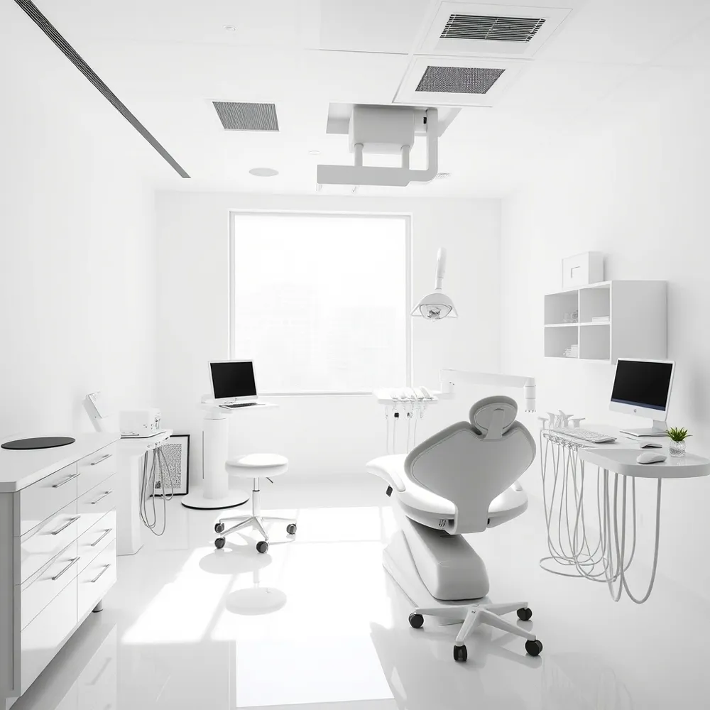
Digital Workflows and Custom Orthodontic Appliances
![]() Modern orthodontic care has transitioned from static, two-dimensional records to dynamic digital models. By integrating Cone Beam Computed Tomography (CBCT) imaging with intraoral scans, clinicians create a comprehensive virtual patient for treatment simulation. This digital replication allows the team at sbdental.com to evaluate tooth roots, bone density, and facial anatomy in a volumetric space, ensuring that every movement is planned with meticulous precision.
Modern orthodontic care has transitioned from static, two-dimensional records to dynamic digital models. By integrating Cone Beam Computed Tomography (CBCT) imaging with intraoral scans, clinicians create a comprehensive virtual patient for treatment simulation. This digital replication allows the team at sbdental.com to evaluate tooth roots, bone density, and facial anatomy in a volumetric space, ensuring that every movement is planned with meticulous precision.
- How is 3D imaging utilized in orthodontics? In orthodontics, 3D imaging and CBCT scans are used to create a virtual patient model. This allows orthodontists to evaluate the relationship between the craniofacial skeleton and soft tissues, enabling highly precise, customized treatment planning that can be interactive and rotated for a complete diagnostic view.
- What are the benefits of 3D-printed orthodontic appliances? 3D-printed orthodontic appliances, such as custom-fit aligners and braces, utilize advanced technology to offer vastly improved precision and personalized treatment plans tailored to your unique anatomy. By integrating digital scans and specialized software, these appliances ensure a superior fit, which leads to enhanced patient comfort and more efficient tooth movement. This streamlined process significantly reduces overall treatment duration, meaning you spend less time in the dental chair and more time enjoying your new smile. Furthermore, the enhanced accuracy of 3D printing helps minimize potential discomfort and fosters better oral hygiene throughout your treatment. At sbdental.com, we embrace these modern solutions to provide your family with a smoother, more supportive, and highly effective orthodontic experience.
When compared to traditional methods that often rely on physical impressions, the digital workflow at sbdental.com offers a clear advantage in accuracy and comfort. While some practices still rely on manual molds, our adoption of 3D data enables the fabrication of appliances that fit your anatomy perfectly from day one. Clinical research published in the Journal of Clinical Medicine highlights that these digital workflows are fundamental to achieving predictable clinical outcomes in complex orthodontic cases.
Safety Protocols and the ALARA Principle
It is completely natural for patients to have questions regarding radiation, as dental X-rays involve ionizing radiation that is often a concern when considering cumulative health effects. To address this, modern dentistry practices like sbdental.com operate under the ALARA principle—which stands for As Low as Reasonably Achievable—to ensure that every diagnostic image is both justified and kept at the lowest possible dose.
Advanced technology, such as 3D CBCT imaging, allows our clinical team to restrict the X-ray beam exclusively to the specific area requiring examination. This precision significantly increases diagnostic accuracy while reducing exposure to the rest of the body compared to older, less focused equipment used by some other clinics. Furthermore, current evidence-based guidelines now emphasize that the use of lead aprons or thyroid collars is no longer necessary, as modern equipment is far more efficient at limiting exposure than traditional protective shielding.
At sbdental.com, our priority is to perform a thorough clinical assessment to determine if an X-ray is necessary for your specific diagnosis. We believe that informed patients make the best decisions for their health, so we are always available to discuss how we maintain these standards. This patient-centered approach ensures you receive the high-quality care you deserve with total peace of mind throughout every phase of your journey.
Your Smile Journey with Advanced 3D Technology
Transitioning to Cone Beam Computed Tomography (CBCT) marks a significant step toward achieving long-term oral health through precision care. By capturing detailed, volumetric data of your unique dental anatomy, this technology allows for more accurate predictive modeling of tooth movement compared to flat, two-dimensional projections. This clarity helps identify complex issues early, reducing the likelihood of unexpected adjustments later in your treatment plan.
At sbdental.com, we leverage these diagnostic tools to provide highly personalized care paths. While some practices rely solely on traditional radiography, which may lack detail on root positioning or bone depth, our commitment to using modern imaging ensures you benefit from a deeper understanding of your treatment needs.
Understanding your own oral structure can be transformative, as it transforms abstract dental concepts into a clear visual guide for your wellness. We invite you to schedule a consultation to discuss how these advanced diagnostic insights at sbdental.com can support your goals. Our team is ready to help you plan your journey toward a lasting, healthy smile with the technology that best fits your specific needs.